Hyperostosis frontoparietalis is a variant of the more common and more well known hyperostosis frontalis interna. As the name suggests, there is benign overgrowth exclusively of the inner table of the frontal bones and parietal bones. Characteristic features include sparing of the midline and outer calvarial surface 1. This is a common.. Hyperostosis frontalis interna (HFI) is a benign entity manifested by bony overgrowth in the frontal endocranial surface. It is most commonly reported incidentally among postmenopausal elderly women. Tracer uptake appearances of HFI can vary on planar bone scans, enabling it to be easily confounded with bone metastases.

Pin on Radiographical Anatomy

Neurocrânio Frontal Parietal Occipital Temporal Esfenoide e Etmoide Anatomia dos

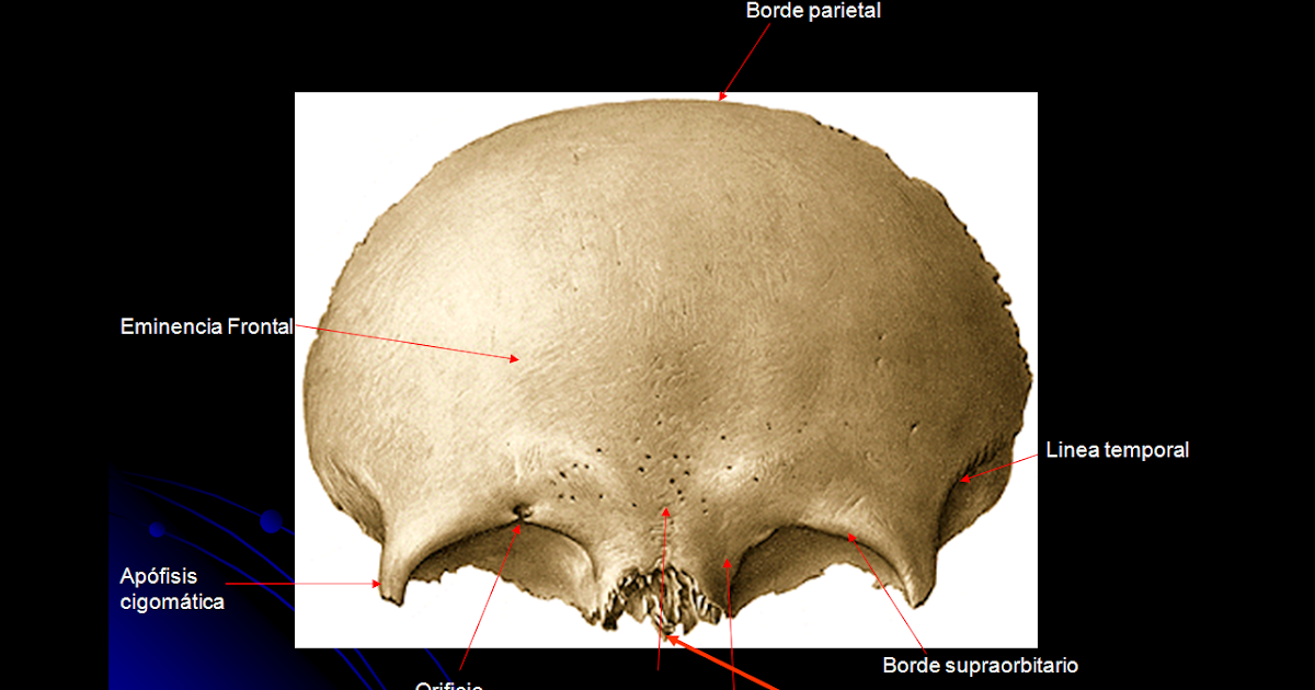
Medicina para Pocos HUESO FRONTAL

Hiperostosis frontal interna; Leontiasis Ósea; Síndrome de

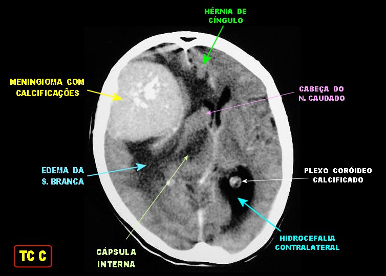
NeupatimagemUNICAMP

Face Leonina na Síndrome Urêmica Brazilian Journal of Nephrology (BJN)

Hiperostose Esquelética Idiopática Difusa (DISH) Dr. Ricardo Teixeira

Hyperostosis Frontalis Interna Cardiology JAMA Neurology JAMA Network

Anatomia Dos Seios Paranasais Vista Lateral Seio Frontal Seio Maxilar My XXX Hot Girl

Dr Balaji Anvekar's Neuroradiology Cases Hyperostosis Frontalis Interna

CT Brain Bone Window Axial Section Shows Bilateral Hyperostosis of the... Download Scientific

Hiperostose frontal interna YouTube

Hiperostose Esquelética Idiopática Difusa (DISH) Dr. Ricardo Teixeira
(PDF) VARIANTES ANATÔMICAS NA CALOTA CRANIANA OBSERVADAS NA TOMOGRAFIA COMPUTADORIZADA ENSAIO

Como Hiperostose frontal interna é diagnosticada?

Quais são os melhores tratamentos para Hiperostose frontal interna?

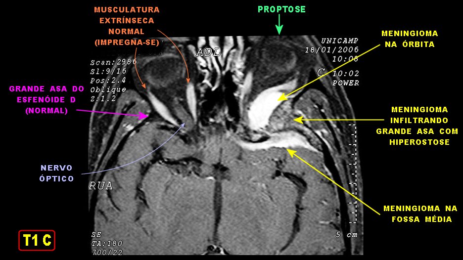
NeupatimagemUNICAMP

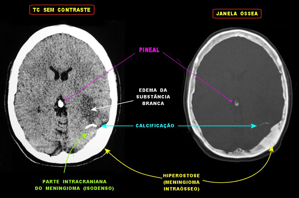
NeupatimagemUNICAMP

¿Cuáles son los síntomas de la Hiperostosis frontal interna?

Hiperostose Esquelética Idiopática Difusa (DISH) Dr. Ricardo Teixeira
Unknown etiology. Hyperostosis frontalis interna is a boney overgrowth of the inner side of the frontal bone of the skull caused by overgrowth of the endocranial surface. It is most often found in women after menopause. It is also associated with hormonal imbalance, being overweight, history of headaches, and neurocognitive degenerative conditions.. The major feature of Hyperostosis Frontalis Interna is excessive growth or thickening of the frontal bone of the head. This excess growth can only be seen in an x-ray. As a result, scientists feel that this condition may be much more prevalent than suspected, but often goes undetected. Many people have no apparent symptoms.